Key points
- Deleting Gata2 from duct-surrounding tissue in mice made the cranial Müllerian duct, which becomes the oviduct, regress.
- The regression did not come from stray anti-Müllerian hormone signaling and may reflect lost NRG1 signaling.
- Gata2 in the duct lining was specifically needed for uterine glands to form in mice.
- Two people with GATA2 deficiency had uterovaginal agenesis or hypoplasia consistent with MRKH syndrome.
The standard textbook story says female embryos hold on to their Müllerian ducts simply because no hormone tells those ducts to disappear. The Müllerian ducts are the embryonic tubes that develop into the oviduct, uterus, cervix and upper vagina. A study in PLOS Genetics now challenges that idea. Working mainly with mice, a team including Shuai Jia and Fei Zhao reports that a gene called Gata2 actively keeps the ducts from breaking down.
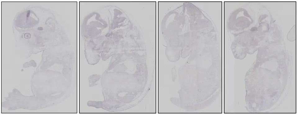
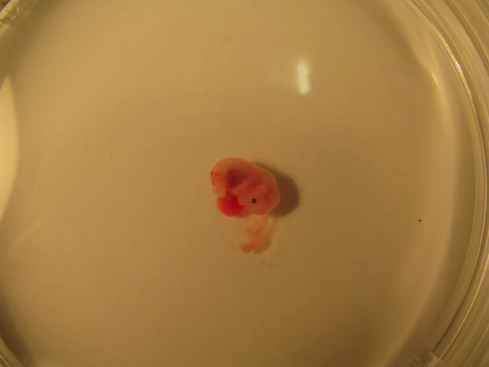
In males, anti-Müllerian hormone (AMH) from the testes makes the ducts regress. The researchers used a genetic tool that targets the mesenchyme, the loose connective tissue that wraps around the ducts, to delete Gata2 there. In those mouse embryos, the cranial end of each duct, the part that forms the oviduct, was initially formed but then degenerated and closed off. From embryonic day 16.5, the upper tract was shorter and less developed than in controls. The team found that the damage was not caused by misplaced AMH signaling. Instead, it may come from losing a signal called NRG1 in that region.
The same mice also had an underdeveloped uterus and a closed-off vagina. When the gene was removed from the duct lining instead, uterine glands specifically failed to form.
The team also describes two people with GATA2 deficiency whose uterus and vagina were missing or underdeveloped. Their condition was consistent with Mayer-Rokitansky-Küster-Hauser (MRKH) syndrome. The authors suggest that GATA2 deficiency could be one genetic cause of such anomalies. The published version is an uncorrected proof.
Why it matters
The authors say the results show that keeping the Müllerian ducts is an active developmental process. They also point to GATA2 deficiency as a possible genetic cause of human Müllerian anomalies such as MRKH syndrome.
What we don't know yet
The mechanism comes from mouse experiments. The human evidence rests on just two individuals. The authors describe the role of NRG1 only as a potential contributor, and the article is still an uncorrected proof.
Terms in this story
- Müllerian ducts
- Paired embryonic tubes that develop into the female reproductive tract, including the oviduct, uterus, cervix and upper vagina.
- Mesenchyme
- Loose, undifferentiated connective tissue in the embryo that sends signals guiding how nearby tissues develop.
- MRKH syndrome
- Mayer-Rokitansky-Küster-Hauser syndrome, a condition in which the uterus and vagina are absent or underdeveloped.
Sources and verification
DexScience articles are produced with AI assistance and an automated fact-check against the linked sources. Spotted an error? Tell us and we will correct it openly.