Key points
- Researchers identified two additional DivIVA domain-containing proteins in *Mycobacterium smegmatis*: Msm0050 and Msm5083.
- Deleting Msm5083, renamed CidIVA, caused membrane defects and disrupted lipid biosynthesis without blocking growth in culture.
- The tuberculosis counterpart Rv1209 gathered at the poles and side wall and mostly bound virulence-related proteins.
- Rv1209 was dispensable in vitro but required for *M. tuberculosis* survival inside a host.
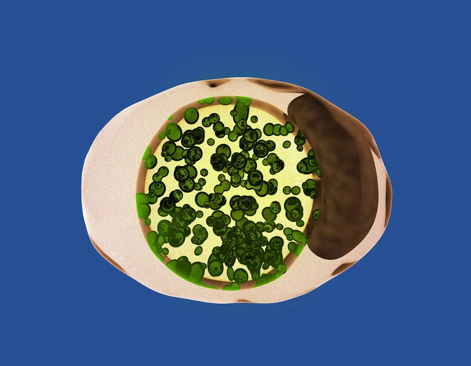
A hunt through mycobacterial genomes for overlooked cell-division machinery has turned up two previously uncharacterized proteins, one of which the researchers have named CidIVA. Writing in PLOS Pathogens, Yogita Kapoor and colleagues report that CidIVA — short for cell envelope integrity determinant DivIVA — helps these bacteria build and maintain the thick, layered coat that surrounds them.
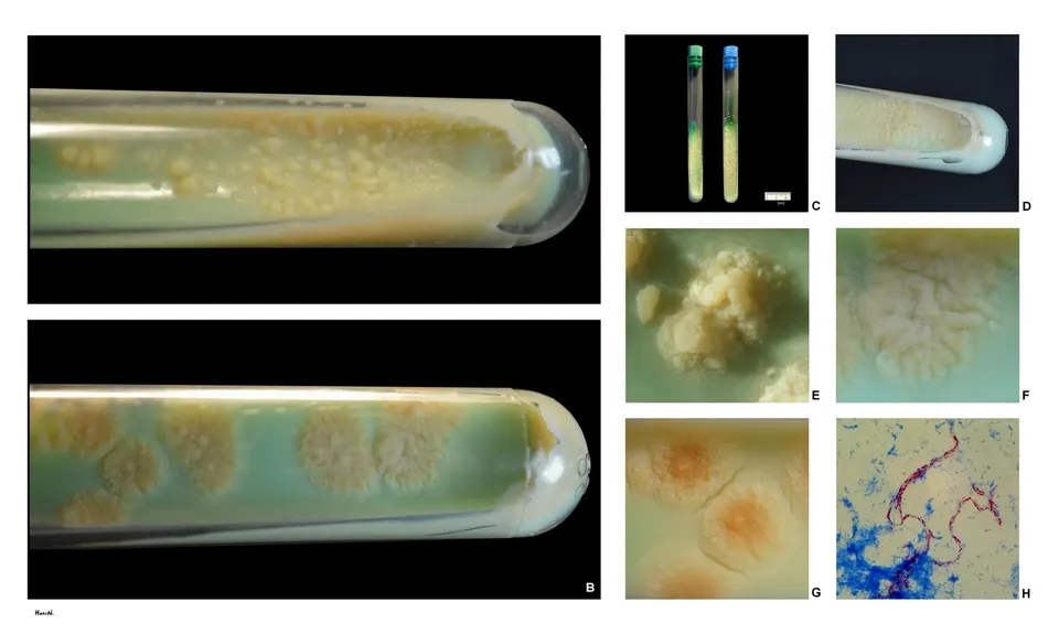
Mycobacteria, the genus that includes the tuberculosis bacterium Mycobacterium tuberculosis, divide differently from laboratory workhorses such as Escherichia coli and Bacillus subtilis. They add new wall material mainly at their tips, and they wrap themselves in an envelope of peptidoglycan, arabinogalactan, mycolic acids, lipids and polysaccharides. Two mycobacterial proteins carrying a so-called DivIVA domain, Wag31 and SepIVA, were already described. Analyzing the mycobacterial proteome, the team found two more in Mycobacterium smegmatis, labeled Msm0050 and Msm5083; an eggNOG database search for homologs of wag31 in M. tuberculosis turned up Rv1209, the counterpart of Msm5083.
Bacteria missing Msm5083 still survived in culture, but they showed pronounced membrane defects and altered lipid production — which is why the authors named the protein for envelope integrity. The protein sits in the membrane and, in interaction studies, associated with other membrane proteins tied to membrane synthesis and upkeep, lipid metabolism and transport.
Its tuberculosis counterpart behaved differently. Rv1209 collected at the poles and along the side wall, and its main partners were proteins linked to virulence. It was not needed for growth in the dish, the researchers say, but it was required for M. tuberculosis to survive inside a host in animal infection experiments. The authors suggest the pair illustrates how members of one protein family can be repurposed for non-overlapping jobs, even in closely related species.
Why it matters
Understanding how the tuberculosis bacterium grows, divides and maintains its protective envelope is a route to finding vulnerable points that drugs might target, and the authors note that mycobacterial growth pathways remain incompletely mapped.
What we don't know yet
Most of the detailed characterization was done in the fast-growing relative *M. smegmatis*, not in *M. tuberculosis*; the host-survival work was in animal infection models, and the paper is posted as an uncorrected proof.
Terms in this story
- DivIVA domain
- A protein module, named after a bacterial cell-division protein, found in several bacteria and linked to organizing growth and division.
- Cell envelope
- The multilayered outer covering of mycobacteria, built from peptidoglycan, arabinogalactan, mycolic acids, lipids and polysaccharides.
- Orthologue
- A gene in one species that corresponds to a gene in another species, inherited from a shared ancestor.
Sources and verification
DexScience articles are produced with AI assistance and an automated fact-check against the linked sources. Spotted an error? Tell us and we will correct it openly.